Facilities and Equipment Used by the Department of Radiological Technology
There is an environment where you can learn practically at the adjacent hospital.
In the medical field, not only knowledge but also how to take appropriate actions based on that knowledge is required. In this department, we have prepared an optimal environment where you can put into practice what you have learned, such as specialized knowledge and skills in handling medical equipment.
University facilities
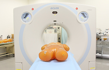
CT device
It is a device that takes a tomographic photograph of the human body using X-rays. It is a device that builds 3D images from the data obtained from the whole body and learns the image processing necessary for clinical practice.
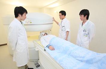
MRI device
It is a device that creates images using magnets and radio waves. Radiological technologists also handle all types of diagnostic imaging equipment that do not use radiation.
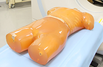
phantom
It is an artificial model for shooting. It is made of a material similar to the human body and is used when taking tomographic photographs and X-rays.
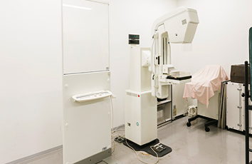
Mammography
Breast x-ray is the official name. It is an indispensable device for diagnosing breast cancer, and you will also learn about care for patients undergoing examination.
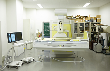
X-ray fluoroscopy device
It is a device that observes the shape and function of organs as an image using a contrast medium such as barium.
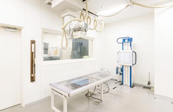
General photography equipment
It is a device that is the basis of medical radiation. We will take pictures of the chest, abdomen, bones, etc. The shooting time is shorter than that of CT or MRI.
Facilities of the attached hospital
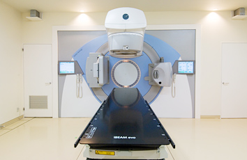
Linac
It is a device that performs treatment by irradiating radiation based on the optimal treatment plan according to the type and spread of the lesion (tumor).
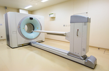
PET-CT device
It is a device that allows you to check the shape and location of cancer as an image in addition to the metabolism and properties of the cancer. It is used to diagnose the presence and spread of various cancers.