Facilities and Equipment Used by the Department of Radiological Technology
Equipped with the latest inspection equipment used in the medical field, we will conduct practical learning
It is fully equipped with various latest inspection equipment used in actual clinical sites. By these equipment, it is possible to acquire biological image information using radiation, magnetism, ultrasound, etc., and to develop knowledge about diagnosis・treatment skills, and practical skills. It also aims to develop accurate knowledge about human body effects such as radiation and electromagnetic waves and safety management.
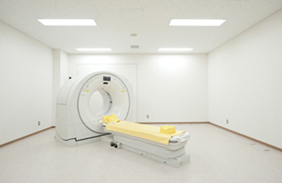
CT device
This is a device that uses X-rays to take cross-sectional images of the human body. Our university uses the latest 16-slice CT scanner, which uses data obtained from the entire body to create 3D images and perform the image processing required for clinical use.
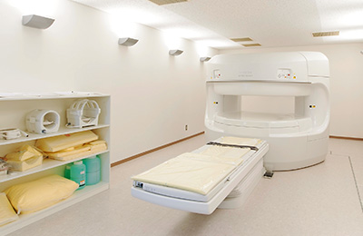
MRI device
This is a device that uses magnets and radio waves to create images, and the device at our university is a permanent magnet type with a magnetic field strength of 0.25 Tesla. radiological technologist also handle all types of imaging diagnostic devices that do not use radiation.
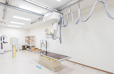
X-ray equipment
This equipment performs the most basic simple imaging with X-rays performed by radiological technologist, which is also called general radiography. Chest, abdomen and bone imaging using this equipment. At our university, we have installed three of these devices.
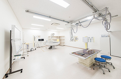
X-ray TV fluoroscope
It is a device that photographs the digestive tract such as the stomach and intestines. Using a contrast medium such as barium, the movement of the digestive tract is displayed as a video in real time, and the captured image is digitally processed.
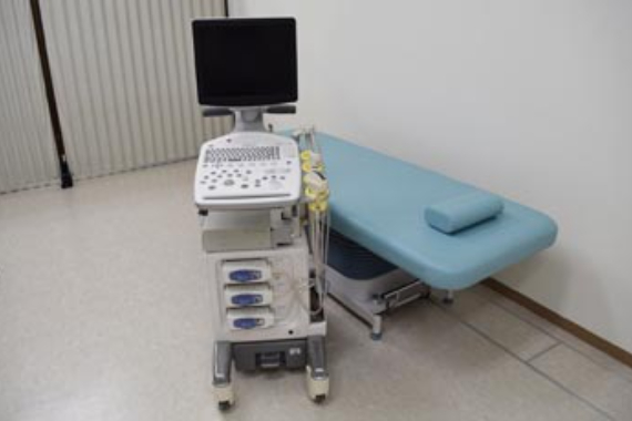
Ultrasonic device
This is a device that uses high-frequency ultrasound that humans cannot hear to take cross-sectional images and examine the condition inside the body (such as the neck, abdomen, heart, and blood vessels). Since it takes images without using X-rays, it is possible to perform the examination without worrying about radiation exposure.
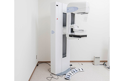
Mammography device
This is an X-ray device specifically designed for the breasts. Also known as mammography, this is the most important method for breast cancer screening in recent years and requires highly detailed technology.
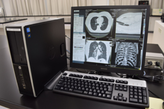
Work station
In order to make images taken by imaging examination devices easier to diagnose, this workstation processes the digital data sent from each device and creates images with high diagnostic capabilities.